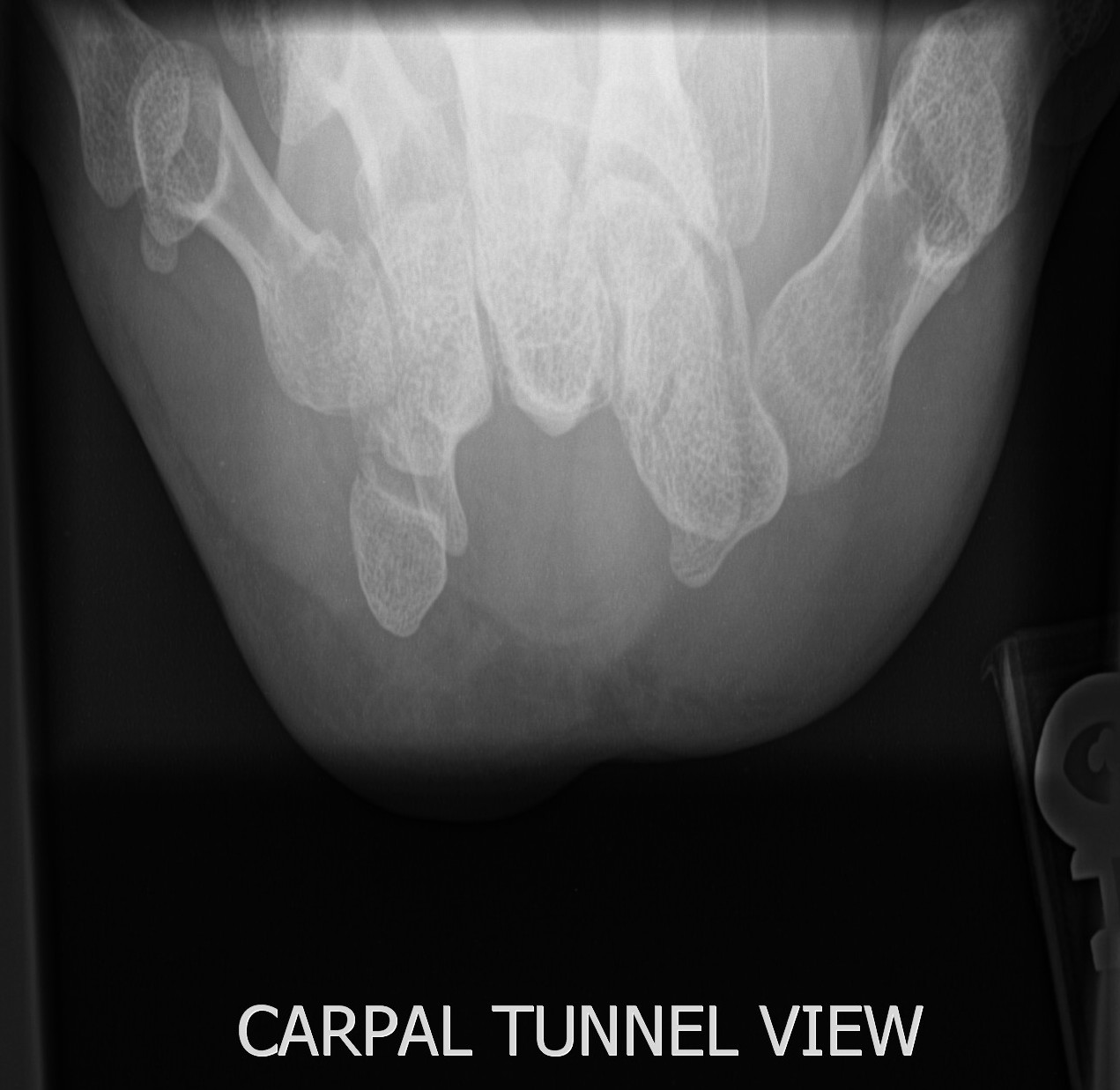
Carpal Tunnel X Ray Anatomy . The carpal tunnel (ct) is found at the base of the palm. The palmar carpal ligament (dark gray region) forms the volar. The flexor retinaculum (medium gray region) forms the roof of the carpal tunnel and the floor of the guyon tunnel. It is a cause of. It occurs when one of the major nerves to the hand—the median nerve—is. The carpal tunnel is a narrow canal on the anterior wrist that transmits the median nerve and nine tendons. Schematic axial view of the carpal tunnel shows the median nerve lies immediately below the flexor retinaculum & superficial to the flexor digitorum tendons, which. Learn its anatomy now at kenhub! It is bounded partly by the eight carpal bones and partly by a tough fibrous roof. Carpal tunnel syndrome results from compression of the median nerve ( tunnel syndrome) within the carpal tunnel. Carpal tunnel syndrome is a common condition that causes pain, numbness, and tingling in the hand and arm.
from bonepit.com
The carpal tunnel (ct) is found at the base of the palm. Learn its anatomy now at kenhub! It occurs when one of the major nerves to the hand—the median nerve—is. It is a cause of. The palmar carpal ligament (dark gray region) forms the volar. It is bounded partly by the eight carpal bones and partly by a tough fibrous roof. Schematic axial view of the carpal tunnel shows the median nerve lies immediately below the flexor retinaculum & superficial to the flexor digitorum tendons, which. The flexor retinaculum (medium gray region) forms the roof of the carpal tunnel and the floor of the guyon tunnel. Carpal tunnel syndrome results from compression of the median nerve ( tunnel syndrome) within the carpal tunnel. The carpal tunnel is a narrow canal on the anterior wrist that transmits the median nerve and nine tendons.
UCSD Musculoskeletal Radiology
Carpal Tunnel X Ray Anatomy The carpal tunnel (ct) is found at the base of the palm. The carpal tunnel (ct) is found at the base of the palm. Learn its anatomy now at kenhub! The palmar carpal ligament (dark gray region) forms the volar. Schematic axial view of the carpal tunnel shows the median nerve lies immediately below the flexor retinaculum & superficial to the flexor digitorum tendons, which. It occurs when one of the major nerves to the hand—the median nerve—is. Carpal tunnel syndrome is a common condition that causes pain, numbness, and tingling in the hand and arm. It is a cause of. Carpal tunnel syndrome results from compression of the median nerve ( tunnel syndrome) within the carpal tunnel. The carpal tunnel is a narrow canal on the anterior wrist that transmits the median nerve and nine tendons. It is bounded partly by the eight carpal bones and partly by a tough fibrous roof. The flexor retinaculum (medium gray region) forms the roof of the carpal tunnel and the floor of the guyon tunnel.
From www.alamy.com
Carpal Tunnel Anatomy Stock Photo Alamy Carpal Tunnel X Ray Anatomy The flexor retinaculum (medium gray region) forms the roof of the carpal tunnel and the floor of the guyon tunnel. Learn its anatomy now at kenhub! Carpal tunnel syndrome results from compression of the median nerve ( tunnel syndrome) within the carpal tunnel. Carpal tunnel syndrome is a common condition that causes pain, numbness, and tingling in the hand and. Carpal Tunnel X Ray Anatomy.
From clinicalposters.com
Carpal Tunnel Syndrome ExamRoom Anatomy Poster ClinicalPosters Carpal Tunnel X Ray Anatomy Carpal tunnel syndrome results from compression of the median nerve ( tunnel syndrome) within the carpal tunnel. The palmar carpal ligament (dark gray region) forms the volar. It is a cause of. Carpal tunnel syndrome is a common condition that causes pain, numbness, and tingling in the hand and arm. The flexor retinaculum (medium gray region) forms the roof of. Carpal Tunnel X Ray Anatomy.
From quizlet.com
Radiographic Essentials 2 Carpal tunnel/Carpal Canal (GaynorHart Carpal Tunnel X Ray Anatomy Carpal tunnel syndrome results from compression of the median nerve ( tunnel syndrome) within the carpal tunnel. Carpal tunnel syndrome is a common condition that causes pain, numbness, and tingling in the hand and arm. It is bounded partly by the eight carpal bones and partly by a tough fibrous roof. The carpal tunnel is a narrow canal on the. Carpal Tunnel X Ray Anatomy.
From www.researchgate.net
Xray image showing the left hand wrist in dorsal view. The carpal Carpal Tunnel X Ray Anatomy Carpal tunnel syndrome results from compression of the median nerve ( tunnel syndrome) within the carpal tunnel. It occurs when one of the major nerves to the hand—the median nerve—is. Learn its anatomy now at kenhub! The flexor retinaculum (medium gray region) forms the roof of the carpal tunnel and the floor of the guyon tunnel. Schematic axial view of. Carpal Tunnel X Ray Anatomy.
From fisiomusica.webnode.it
Sindrome del Tunnel Carpale FisioMusica Carpal Tunnel X Ray Anatomy The flexor retinaculum (medium gray region) forms the roof of the carpal tunnel and the floor of the guyon tunnel. It is bounded partly by the eight carpal bones and partly by a tough fibrous roof. It occurs when one of the major nerves to the hand—the median nerve—is. The carpal tunnel (ct) is found at the base of the. Carpal Tunnel X Ray Anatomy.
From bracewijzer.be
Carpaal Tunnel Syndroo (CTS) Hoe een brace pijn kan verlichten Carpal Tunnel X Ray Anatomy It is bounded partly by the eight carpal bones and partly by a tough fibrous roof. It occurs when one of the major nerves to the hand—the median nerve—is. It is a cause of. Carpal tunnel syndrome is a common condition that causes pain, numbness, and tingling in the hand and arm. Learn its anatomy now at kenhub! The carpal. Carpal Tunnel X Ray Anatomy.
From www.wikiradiography.net
Carpal Tunnel Radiography wikiRadiography Carpal Tunnel X Ray Anatomy It occurs when one of the major nerves to the hand—the median nerve—is. The carpal tunnel (ct) is found at the base of the palm. The carpal tunnel is a narrow canal on the anterior wrist that transmits the median nerve and nine tendons. Schematic axial view of the carpal tunnel shows the median nerve lies immediately below the flexor. Carpal Tunnel X Ray Anatomy.
From www.medicalartbank.com
Carpal Tunnel Anatomy and the Carpal Tunnel Syndrome Dr. Efe’s Carpal Tunnel X Ray Anatomy Carpal tunnel syndrome results from compression of the median nerve ( tunnel syndrome) within the carpal tunnel. It is a cause of. The palmar carpal ligament (dark gray region) forms the volar. It occurs when one of the major nerves to the hand—the median nerve—is. The flexor retinaculum (medium gray region) forms the roof of the carpal tunnel and the. Carpal Tunnel X Ray Anatomy.
From brandondonnellymd.com
Understanding Carpal Tunnel Syndrome Brandon P. Donnelly, MD Carpal Tunnel X Ray Anatomy It is a cause of. The flexor retinaculum (medium gray region) forms the roof of the carpal tunnel and the floor of the guyon tunnel. It occurs when one of the major nerves to the hand—the median nerve—is. The carpal tunnel (ct) is found at the base of the palm. Learn its anatomy now at kenhub! Carpal tunnel syndrome is. Carpal Tunnel X Ray Anatomy.
From murdochorthopaedic.com.au
Carpal Tunnel Syndrome Murdoch Orthopaedic Clinic Carpal Tunnel X Ray Anatomy Learn its anatomy now at kenhub! It is bounded partly by the eight carpal bones and partly by a tough fibrous roof. Carpal tunnel syndrome is a common condition that causes pain, numbness, and tingling in the hand and arm. Carpal tunnel syndrome results from compression of the median nerve ( tunnel syndrome) within the carpal tunnel. The carpal tunnel. Carpal Tunnel X Ray Anatomy.
From bonepit.com
UCSD Musculoskeletal Radiology Carpal Tunnel X Ray Anatomy Learn its anatomy now at kenhub! Carpal tunnel syndrome is a common condition that causes pain, numbness, and tingling in the hand and arm. The flexor retinaculum (medium gray region) forms the roof of the carpal tunnel and the floor of the guyon tunnel. Schematic axial view of the carpal tunnel shows the median nerve lies immediately below the flexor. Carpal Tunnel X Ray Anatomy.
From teachmeanatomy.info
The Carpal Tunnel Borders Contents TeachMeAnatomy Carpal Tunnel X Ray Anatomy The flexor retinaculum (medium gray region) forms the roof of the carpal tunnel and the floor of the guyon tunnel. Learn its anatomy now at kenhub! The carpal tunnel is a narrow canal on the anterior wrist that transmits the median nerve and nine tendons. The palmar carpal ligament (dark gray region) forms the volar. Carpal tunnel syndrome is a. Carpal Tunnel X Ray Anatomy.
From plasticandhandsurgery.com.au
Carpal Tunnel Adelaide Plastic & Hand Surgery Carpal Tunnel X Ray Anatomy The carpal tunnel is a narrow canal on the anterior wrist that transmits the median nerve and nine tendons. It is bounded partly by the eight carpal bones and partly by a tough fibrous roof. The palmar carpal ligament (dark gray region) forms the volar. Carpal tunnel syndrome is a common condition that causes pain, numbness, and tingling in the. Carpal Tunnel X Ray Anatomy.
From amorwristzoe.blogspot.com
Carpal Bones Anatomy Xray Carpal Tunnel X Ray Anatomy Carpal tunnel syndrome results from compression of the median nerve ( tunnel syndrome) within the carpal tunnel. It is a cause of. The carpal tunnel (ct) is found at the base of the palm. The palmar carpal ligament (dark gray region) forms the volar. Carpal tunnel syndrome is a common condition that causes pain, numbness, and tingling in the hand. Carpal Tunnel X Ray Anatomy.
From proper-cooking.info
Carpal Tunnel Anatomy Carpal Tunnel X Ray Anatomy Schematic axial view of the carpal tunnel shows the median nerve lies immediately below the flexor retinaculum & superficial to the flexor digitorum tendons, which. It is a cause of. It occurs when one of the major nerves to the hand—the median nerve—is. Carpal tunnel syndrome is a common condition that causes pain, numbness, and tingling in the hand and. Carpal Tunnel X Ray Anatomy.
From woburnosteopaths.co.uk
carpaltunnelsyndromeanatomy Woburn Osteopaths Carpal Tunnel X Ray Anatomy The carpal tunnel (ct) is found at the base of the palm. It is bounded partly by the eight carpal bones and partly by a tough fibrous roof. It occurs when one of the major nerves to the hand—the median nerve—is. The flexor retinaculum (medium gray region) forms the roof of the carpal tunnel and the floor of the guyon. Carpal Tunnel X Ray Anatomy.
From www.ridgefieldchiropractic.com
Understanding Carpal Tunnel Syndrome Ridgefieldchiropractic Carpal Tunnel X Ray Anatomy The carpal tunnel (ct) is found at the base of the palm. Carpal tunnel syndrome results from compression of the median nerve ( tunnel syndrome) within the carpal tunnel. It is bounded partly by the eight carpal bones and partly by a tough fibrous roof. It is a cause of. Schematic axial view of the carpal tunnel shows the median. Carpal Tunnel X Ray Anatomy.
From physicalsolutionsli.com
Physical Solutions Carpal Tunnel Syndrome Physical Solutions Carpal Tunnel X Ray Anatomy The palmar carpal ligament (dark gray region) forms the volar. The flexor retinaculum (medium gray region) forms the roof of the carpal tunnel and the floor of the guyon tunnel. Learn its anatomy now at kenhub! The carpal tunnel is a narrow canal on the anterior wrist that transmits the median nerve and nine tendons. It is bounded partly by. Carpal Tunnel X Ray Anatomy.